

In a darkened lab at MIT, a zebrafish larva barely longer than a grain of rice is swimming in a dish, and its entire brain is lighting up like a tiny galaxy. Every flash is a neuron firing. A microscope adapted by engineers in Ed Boyden's Synthetic Neurobiology Group can now watch those flashes across the whole brain, 200 times every second. For the first time, neuroscientists can see an entire brain computing in something close to real time.
The work, published in Nature Methods (DOI: 10.1038/s41592-026-03179-7), solves a problem that has frustrated brain researchers for decades. Neurons think in electricity: millisecond voltage spikes that race through vast networks. But the tools for watching large volumes of brain tissue have been stuck at the speed of a slow-motion replay, until now.
The slow camera problem
The workhorse of brain imaging is calcium imaging. After a neuron fires, calcium ions flow into the cell, and fluorescent dyes report that influx. It is a useful proxy, but a sluggish one. Calcium signals unfold over seconds or even minutes, far too slow to capture individual spikes. "Calcium imaging inherently is very slow," says lead author Jie Zhang, "so you're talking about imaging activity on the order of seconds or even minutes." The brain does its most interesting work on a timescale thousands of times faster.
The alternative is voltage imaging: measuring the electrical signal directly. Genetically encoded voltage indicators are fluorescent proteins engineered into neurons, and they light up when the neuron fires. It is a direct readout of the computation itself, not an echo of it. But until now, voltage imaging had been confined to small, localized populations of neurons, a close-up where the science needed a wide shot.
The MIT team set out to break the tradeoff between speed and coverage. Neurons distributed across the brain coordinate on millisecond timescales to generate behaviors and computations, explains lead author Zeguan Wang, who designed the microscope. To understand those principles, researchers need to observe all of them at once, across the whole brain, so that no important participant neurons are missed.
How they sped it up
Enjoying this story?
Get the five most important stories in tech, every morning. Free.
The instrument is a modified light sheet microscope, a device that illuminates a thin slice of tissue with a sheet of laser light, then builds 3D volumes by stacking slices. Scanning a full volume used to take far too long to catch neural impulses at single-cell resolution.
Two upgrades changed that. The researchers dramatically increased the camera's image acquisition speed and boosted the scanning speed with a technique called remote refocusing. Together, the changes pushed the system to scan the entire larval zebrafish brain 200 times per second, or once every five milliseconds.
To make the firing visible, the team engineered the larvae's neurons to express a voltage indicator called Positron2-Kv. The indicator did not reach every neuron: roughly one quarter produced usable signals. But that quarter, distributed throughout the brain, was enough to observe single spikes and rapid bursts rippling across entire regions.
All of the parts of the brain are connected together, so if you want to truly understand the brain, you have to understand how all the neurons work together as an emergent whole.
That is Ed Boyden, the Y. Eva Tan Professor in Neurotechnology at MIT and the study's senior author, and it is also the argument for the machine. The great open question, he says, is how neurons work together as a network, and this may be the first tool that can actually watch it happen.
What the camera saw
Watching a Brain Think: The Speed Leap
Temporal resolution of brain imaging methods.
Note: Schematic comparison for illustrative purposes.
With the fish at rest, the microscope picked out single voltage spikes from individual neurons, along with rapid bursts of spikes, the kind of fine-grained electrical chatter that calcium imaging smears into an indistinguishable glow.
Then the researchers shone ultraviolet light on the larvae. The response started in the optic tectum, the region that receives and processes visual input from the retina, and the team watched activity propagate across the tectum from one side to the other. It was a wave of computation, caught mid-flight, in a brain smaller than a sesame seed.
Elsewhere, the camera caught something stranger: sequences of spontaneous activity rippling through neurons in the cerebellum and hindbrain with no stimulus at all, the brain's background processes unfolding on their own schedule.
Why drug hunters are watching
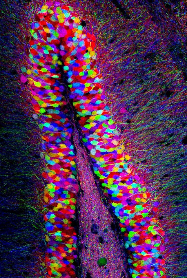
The paper itself is a methods advance. The renewed attention, with neuroscience trade press covering the findings this week, is about what the method enables. Preclinical neuroscience has a measurement gap: candidate drugs for neurological and psychiatric conditions are usually evaluated with slow or highly localized readouts. A brain-wide, millisecond-resolution readout changes what a screening program can ask.
Larval zebrafish are already a standard model for compound screening: small, transparent, and cheap to raise in the thousands. Pairing them with whole-brain voltage imaging could give pharmaceutical researchers a direct window into how a compound reshapes distributed circuit activity, rather than a sluggish calcium echo. It would not replace mammalian studies, but it could become a fast first filter: does this molecule change how a living brain computes, and where?
For neurotechnology developers, the platform is a hypothesis engine. Mapping which neurons fire first, where a sequence begins, and how activity recruits the rest of the network turns vague questions about circuit function into concrete, testable maps.
What comes next
The researchers are candid about the limits. Only a fraction of neurons produced clean signals, and the team wants that fraction to grow, along with the speed and resolution. The bigger leap is biological: extending the technique from zebrafish larvae to mice, whose brains are orders of magnitude more complex and opaque.
If that works, the implications widen. Whole-brain voltage imaging in a mammal could offer new ways to generate hypotheses about what the brain does during specific behaviors, or even during quiet states like daydreaming, the kind of background activity this method already glimpsed in a fish.
Neuroscience has spent a century inferring what the brain does from slow proxies and tiny peepholes. Now it has a camera that keeps up with the electricity. The first subject was a fish. It will not be the last.
164 Comments